
Characterization of Bacterial Communities in Venous Insufficiency Wounds by Use of Conventional Culture and Molecular Diagnostic Methods
ABSTRACT
Microbial infections delay wound healing, but the effect of the composition of the wound microbiome on healing parameters is unknown. To better understand bacterial communities in chronic wounds, we analyzed debridement samples from lower-extremity venous insufficiency ulcers using the following: conventional anaerobic and aerobic bacterial cultures; the Ibis T5000 universal biosensor (Abbott Molecular); and 16S 454 FLX titanium series pyrosequencing (Roche). Wound debridement samples were obtained from 10 patients monitored clinically for at least 6 months, at which point 5 of the 10 sampled wounds had healed. Pyrosequencing data revealed significantly higher bacterial abundance and diversity in wounds that had not healed at 6 months. Additionally, Actinomycetales was increased in wounds that had not healed, and Pseudomonadaceae was increased in wounds that had healed by the 6-month follow-up. Baseline wound surface area, duration, or analysis by Ibis or conventional culture did not reveal significant differences between wounds that healed after 6 months and those that did not. Thus, pyrosequencing identified distinctive baseline characteristics of wounds that did not heal by the 6-month follow-up, furthering our understanding of potentially unique microbiome characteristics of chronic wounds.
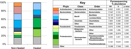
Figure 1
Taxa identified by pyrosequencing. The stacked graph illustrates the relative abundance of each taxon identified by pyrosequencing from the data from baseline samples grouped as nonhealed (NH) or healed (H) based on whether the patient was healed at 6 months (color coded according to the key). The table shown in the key details the total percent abundance of each taxon in the nonhealed versus healed wounds, showing significantly more Actinomycetales in nonhealed wounds and Pseudomonadaceae in healed wounds.
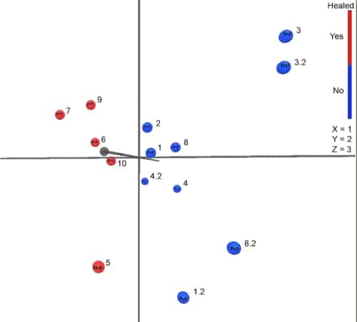
Figure 2
Principle component analysis for wound samples. Principle component analysis plot showing separation of wound samples from patients nonhealed (blue) and healed (red) at 6 months. All samples were included in this analysis, with second samples as x.2, where x is the patient number.